
As we did with the earliest commercial silicon probes in 2004, NeuroNexus is again advancing the field. We are now offering SiNAPS Pixel fully-integrated silicon CMOS probes alongside a complete turn-key recording system to provide you ultra-high density (UHD) neural recording in a cost-effective solution. The probes and system are available now, with promotional pricing and support to early adopters.
NeuroNexus SiNAPS probes use the pioneering new Active Pixel Sensor (APS) technology in which active circuits for signal amplification, low-pass filtering, and multiplexing read-out are located directly underneath each electrode-pixel. The probes are used with a cost-effective, turn-key recording system that runs Radiens™ Allego software, featuring automated mapping, monitoring, and visualization of all electrode sites simultaneously and more.
NeuroNexus is partnering with Corticale to commercialize this exciting new option for high-performance, cost-effective ultra-high density (UHD) neural recording.
The video displayed above was captured during the SiNAPS data collection process at Scott Pluta Lab using a multi-probe manipulator (MPM) system.
The SiNAPS recording system raises the standard for high-definition brain and cardiac mapping by integrating density, resolution, and automation. It provides unparalleled clarity at every level of complexity. Please see below for the three key benefits that distinguish the SiNAPS recording system.
Quantity of Data: More Data. Better Outcomes
With its high point density, the SiNAPS recording system minimizes the need for interpolation between annotated points. As a result, you can visualize propagation patterns more accurately and identify small gaps and areas of interest with greater precision, leading to improved outcomes.
The SiNAPS recording system stands out for its:
Quality of Data: Higher-Quality Data. Better Decision-Making
The SiNAPS recording system boasts high-density coverage, resulting in ultra-clear contact single-unit signals. Utilizing the Radiens Allego Software, the system’s HD maps provide exceptional detail for identifying gaps in locate channels, characterizing complex substrates, and visualizing the propagation of intricate circuits.
What sets SiNAPS apart:
Speed of Data: Data Accuracy and Speed Bring Unparalleled Clarity
The Radiens Allego software sets itself apart from other systems that rely on manual point-by-point annotation by enabling continuous, rapid, and automatic mapping. This innovative approach results in increased consistency, speed, and accuracy.
The SiNAPS recording system, which utilizes the Radiens Allego software, features a novel annotation algorithm that has been shown to deliver an impressive accuracy rate of 99.98%. Moreover, the system collects thousands of relevant data points in just minutes, dramatically reducing map-acquisition time and allowing for more efficient data analysis.
Additional Advantages:
RMS Noise
6.5 μV (300-7500 Hz)
In-pixel amplifier
46 dB (DC-4 kHz)
Power Consumption
<6 μW/electrode-pixel
Sampling Frequency
20 ksample/s
Electrode Size
14×14 μm2
Electrode/Channels
256, 1024
Electrode Pitch
29 μm
Shank Spacing (center-to-center)
560±2 μm (for 4-shank probe)
Electrode Material
Platinum
Shank Length
5656± 60 μm; active length: 3768± 1 μm
Shank Width
88±2 μm
Shank Thickness
50±5 μm


Below is a recording obtained from a Radiens Allego of a head-fixed (awake) mouse using a SiNAPS four-shank probe with 1024 simultaneously recorded channels.
During the week of May 1st, a small team of NeuroNexus engineers and scientists visited a top systems neuroscience lab in New York to initiate a collaborative study using our new SiNAPS Pixel probes to investigate neural circuit dynamics with unprecedented spatiotemporal resolution. While this study is ongoing, we’re pleased to highlight some early findings:
We have reached a significant milestone in our efforts to create increasingly powerful, user-friendly, and affordable electrophysiology tools and solutions that will drive progress in systems neuroscience.
Perhaps the most exciting aspect of this work is that these probes and solutions are available NOW for you to incorporate into your work. Please contact us for more information.
Here is a 3D visualization of neural activity captured using a SiNAPS 1024-channel probe, shown in a brain-centered view.
Presented below are images showcasing acute recordings obtained from a single- and 4-shank SiNAPS probe that was implanted in an anesthetized rodent.

Displayed at the top is an example of in vivo neural activity recordings obtained through a SiNAPS probe implanted in the brain of an anesthetized rat, with the probe’s location specified in the top-left inset. The curves signify the full-band bioelectrical signals recorded from the somatosensory cortex to the hippocampus for a time period of 7 seconds. A total of 320 electrodes were employed to record physiologically-related signals, with only a subset of 110 electrodes (pitch of 28 µm) displayed to avoid overwhelming the visualization. The bottom panel presents a detailed view of a single raw neural trace recorded in the hippocampus, both in its original form and after being filtered offline in the LFP (1–300 Hz) and AP (300–5000 Hz) frequency bands. Further details can be found by following this link.

In vivo recordings were conducted with a multi-shank SiNAPS probe. A. For each shank, a representative subset of 100 ms of broadband neural signals from the available 1024 channels was collected and displayed. B. A raster plot of single unit activity (n = 219) recorded from the entire probe was presented, with each color indicating the macrostructure from which a specific neuron was obtained. C. Additionally, an example of bandpass filtered (300-5000 Hz) neural data collected from a portion of one of the shanks (red area composed of 108 neighboring consecutive channels) of the probe depicted in (A) was shown, along with the waveforms of a subset of units recorded from those channels on the right side of the image. Further details can be found by following this link.

The above image display single-unit light-evoked responses using a 4-shank probe. A. Representation of the probe features spike waveforms of the isolated single-unit responses, with colored dots indicating the location of single units on the probe. B. Examples of the spike rasters and peri-stimulus histograms are shown for optogenetic stimulation of six isolated single units, with the color coding indicating the relative position of the isolated single units on the probe. Light-blue rectangles indicate the period in which the optostimulation is on.

The image above shows the optogenetic LFP light-evoked responses. A. 4-shank probe was positioned and superimposed onto an approximate mouse atlas coronal section (Allen Mouse Brain Atlas). Representative 50ms portions of LFP responses collected along the probe shanks after the light stimulus onset are shown in black curves. A false-color map represents the interpolated peak-to-peak values of the low-frequency responses over the probe’s two-dimensional plane, with the estimated fiber tip position projected on the probe’s coronal plane highlighted by a blue circle. B. Example of the broadband signals recorded from five neighboring channels sampled from the four different shanks is presented, with the blue rectangle indicating light stimulation and the yellow rectangle showing the time used to generate the false-color map. Further details can be found by following this link.
References:
Available Options:
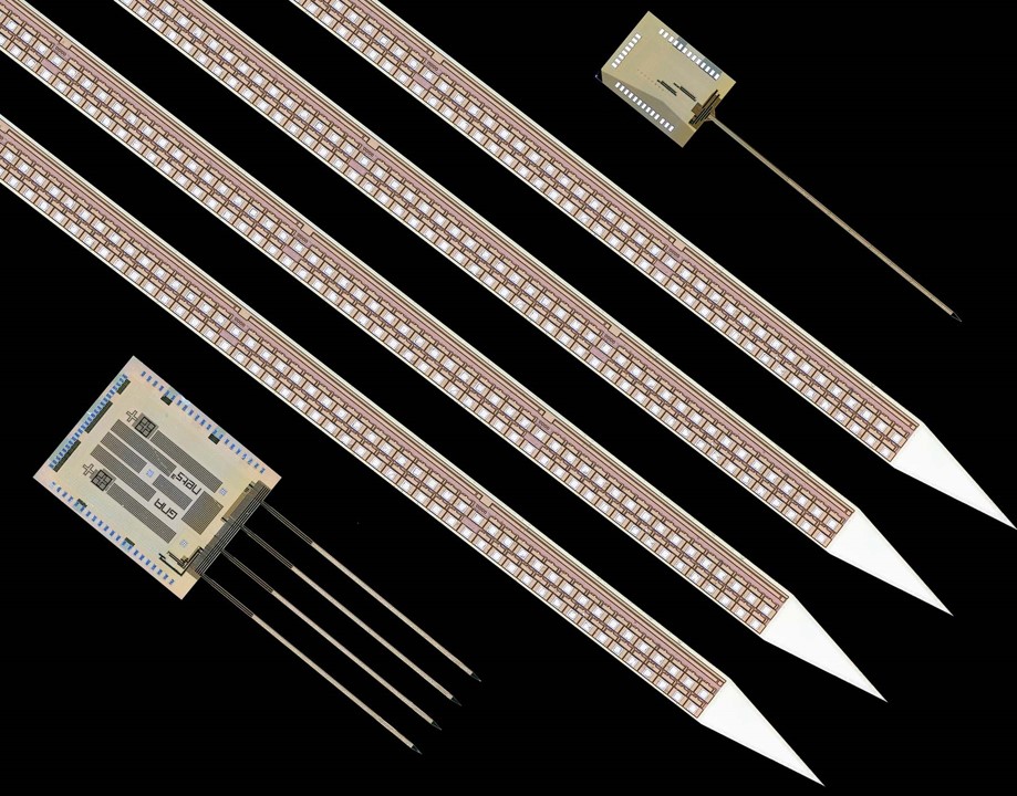
Electrodes: 256 & 1024 recording sites

Available Packages:
System Requirements
Check SiNAPS Knowledge Base for a brief overview of the SiNAPS system operation and SiNAPS electrode calibration.

This image provides an example of in vivo neural activity recording using a SiNAPS probe. A) The architecture features a single shank with 256 SiNAPS probes implanted into the mouse brain. B) An averaged spike waveform from each individual site is shown as an example. C) A subset of 32 sites is shown in a close-up view that has been off-line filtered in the local field potential (LFP) frequency band (1-300 Hz). D) The raster time series of all sites off-line filtered in the action potential (AP) frequency band (300-5000 Hz) are displayed.

SiNAPS optogenetic packages come with an attached single optical fiber for 1-shank 256ch and up to four optical fibers for 4-shank 1024ch designs. The placement of fibers on the 1024 design is unrestricted. Whether one intends to include a single fiber or more, they have the flexibility to position it between shanks or on any preferred shank location.

Please note that we provide flat fibers with 50 µm/62.5 µm, 105 µm/125 µm, or 200 µm/220 µm (inner diameter/outer diameter) options, as well as Lambda fibers with 105 µm/125 µm, 200 µm/225 µm, or 200 µm/230 µm (inner diameter/outer diameter) for SiNAPS Opto packages. For further information, visit the Optogenetics section.

SiNAPS Webinar Recording is Now Live!
The webinar recording is now available and ready for viewing! We are excited to share this valuable content with you. Please feel free to watch at your convenience.
