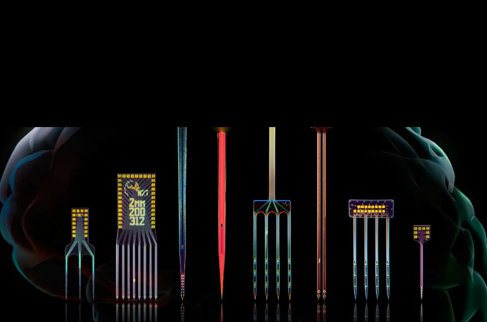
Electrodes for brain, spinal cord, nerve, and heart
Silicon probes and thin-film grids in more than 260 designs, with 4 to 1,024+ sites.
5,200+ papers
Peer-reviewed papers that cite NeuroNexus electrodes
1,000+ labs
Research institutions on six continents
260+ designs
210+ silicon probe designs and 50+ thin-film grid designs, each with a datasheet
4 to 1,024+ sites
From a four-site Q-Trode to active-pixel SiNAPS probes
Browse by requirement
Four questions narrow the catalog: penetrating or surface, where you record, what the device does, and what you measure.
1 · Penetrating or surface
Silicon probes penetrate the brain, spinal cord, nerves, ganglia, or heart. Grid electrodes record from the surface of the cortex, the skull, the heart, or a peripheral nerve.
Silicon probes
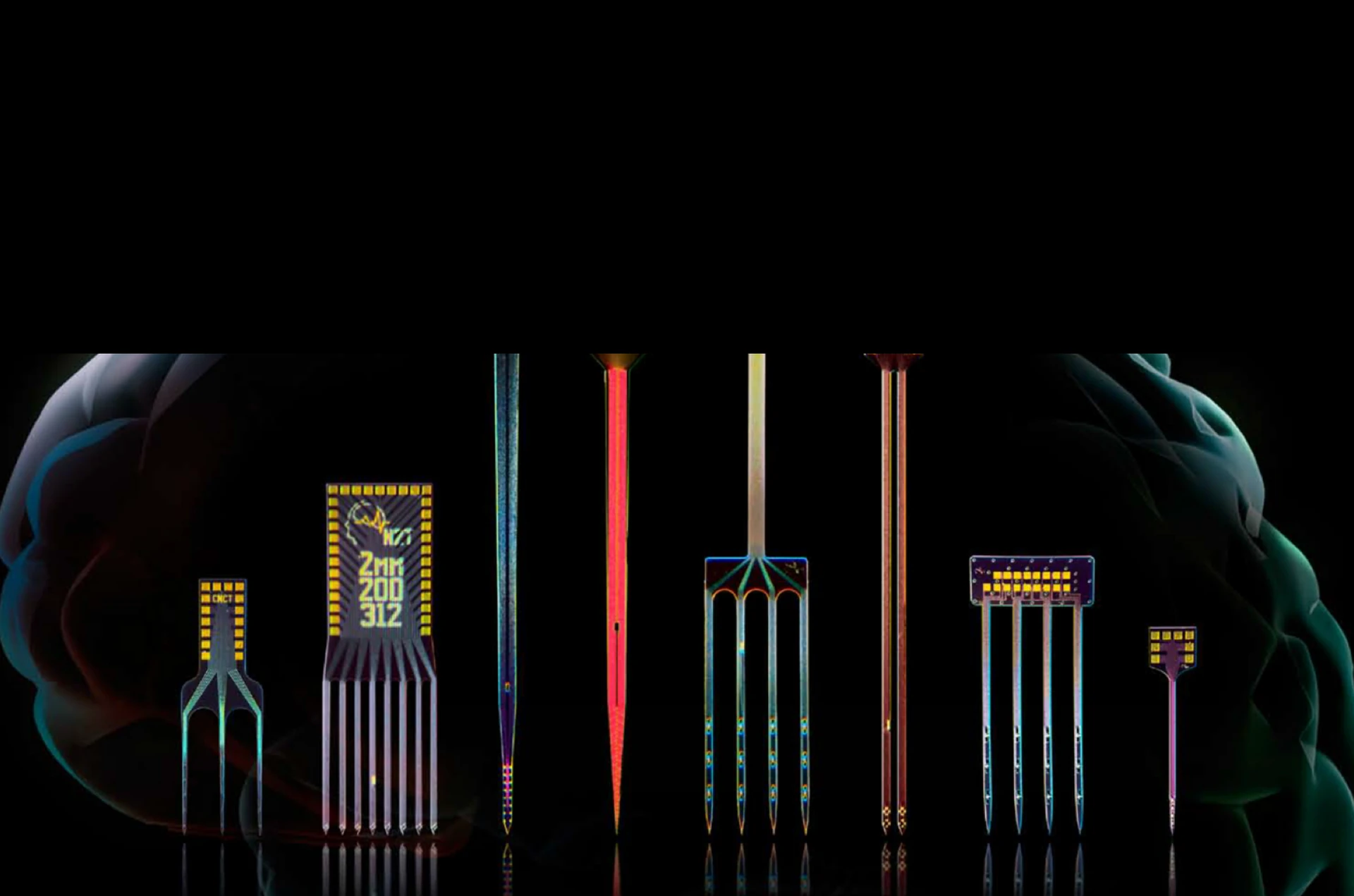
A silicon probe, also called a silicon neural probe or silicon microelectrode array, is a penetrating neural probe made by thin-film microfabrication on a silicon substrate.
- Vector Array · Fusion · Matrix Array · High-Density Series (SiNAPS)
- A-Series · Q-Trode · Drug Delivery · DBS
Grid electrodes
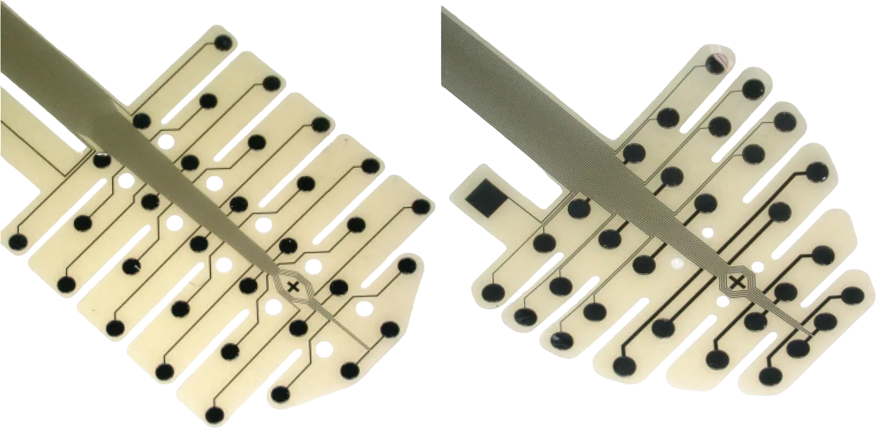
A grid electrode is a flexible array of electrode sites in a precise grid layout that is placed on a tissue surface, where it records field potentials or delivers stimulation without penetrating the tissue.
- ECoG · EEG · cardiac surface · nerve cuff
2 · Target
Anatomy sets the first constraints: depth, access, and how much the tissue moves.
3 · Modality
Modality sets the active elements: sites alone, stimulation-rated sites, a fiber beside the sites, or a fluidic channel.
4 · Signal
Site size and spacing follow the signal. Small, closely spaced sites isolate spikes, and larger sites record LFP, ECoG, EEG, and ECG.
Common to every electrode line
Recording, electrical stimulation, optogenetics, and targeted drug delivery.
Multiple connector and cable packages, by preparation and headstage.
NeuroNexus systems and software, and common third-party headstages and acquisition systems.
Finding the right electrode
Three ways to narrow 260+ designs to the one that fits.
Filter by species, target, signal, site count, and shank geometry, with stock shown per design.
Six questions that select an electrode from the target, measurement, duration, subject, and recording system.
Species, target, signal, and channel count are enough. An Application Scientist replies with designs that fit, or with a custom design if none does.
NeuroNexus electrodes record into Radiens through any compatible headstage and acquisition system. Radiens software is free to start.
Published work with NeuroNexus electrodes
More than 5,200 peer-reviewed papers cite NeuroNexus electrodes. Bioz indexes them with their figures, searchable by technique.
Not sure which fits?
Tell us the species, the target, and the channel count. An Application Scientist will reply with designs that fit.
Choosing an electrode — FAQ
How do the three browse axes relate?
They are orthogonal — one probe can appear under a target, a modality, and a signal. Start from whichever constraint is fixed first, then narrow with the others.
Can one probe span multiple modalities?
Yes. Optoelectrodes combine recording with optical stimulation; drug-delivery probes add a fluidic channel. A device can belong to several modality collections.
Which axis should I start with?
Start from the constraint you cannot change — usually the anatomical target or the signal band — then filter by the others in the webstore.
Where do I see specs and pricing?
Every collection card opens the webstore filtered to that collection, where you can compare channel counts, site geometry, and configuration.
Why does silicon probe thickness matter?
Substrate thickness sets bending stiffness, the tissue the shank displaces, handling, and how deep a probe can be driven without buckling. Catalog designs come at 15 and 50 µm: the thinner substrate displaces less tissue and bends more; the thicker holds a straight trajectory on long insertions. For deep targets in large animals the Vector Array mounts a 10 to 15 mm silicon array on a rigid 70 or 110 mm support body. The process also reaches 5 µm, a thickness at which less tissue encapsulation has been reported (Seymour and Kipke, 2007); that is a capability discussed per design, not a catalog line.