EEG Recording in Mice: Electrode Options
Screw electrodes, a skull-surface thin-film grid, or a wireless headmount: what each gives and costs in a mouse, and when the 30-site grid is the answer.
8 min read
Updated October 28, 2026

NeuroNexus · neuronexus.com · Resources
EEG in a mouse is recorded from the skull surface, and the electrode choice is between a few bone-screw electrodes, a thin-film grid of 16 to 32 sites aligned to bregma, and the system that carries the signal off the head. NeuroNexus makes the grid: 12 µm polyimide with 500 µm platinum sites, placed with no craniotomy and held for weeks to months. This guide sets out what each option gives and costs, and when the grid is the answer.
Research use only. NeuroNexus products and the procedures described here are for non-clinical research use, and must not be used in human or veterinary medical procedures.
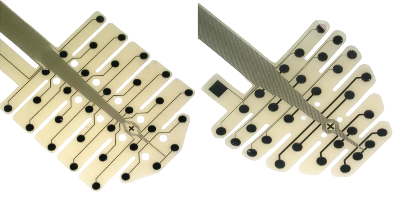
Figure 1. Two mouse EEG grids from the EEG Series: 12 µm polyimide, 500 µm platinum sites laid out to the mouse skull, with the bregma crosshair.
Which electrode records EEG in a mouse?#
Every mouse EEG electrode sits outside the brain. The skull and scalp spread the signal, so what reaches the electrode is the summed rhythm of the cortex beneath it: sleep and wake state, spectral power by band, seizures, evoked responses, and the coherence between regions. No skull-surface electrode records spikes, and none of the three options below changes that. What they change is how many places on the skull are sampled at once, how reproducibly those places are chosen, and what the surgery costs the animal.
| Bone-screw electrodes | Thin-film EEG grid | Wireless headmount | |
|---|---|---|---|
| Channels | one per screw; two to four in practice | 16, 30 or 32 on one film | few; set by the transmitter |
| Placement | each screw drilled and placed by hand | one film, crosshair aligned to bregma | the electrodes it is wired to |
| Surgery | burr holes for each screw | no craniotomy; screws as anchors and reference | as its electrodes |
| Between animals | positions vary with the hand | the same site is the same region in every animal | as its electrodes |
| Tether | yes | yes, through a 32-channel headstage | none |
What does a screw electrode give?#
A stainless-steel screw in a burr hole is the traditional mouse EEG electrode, and for one or two channels it remains the simplest: a seizure count, a sleep score, a single cortical rhythm. Its limits are the two things a screw cannot do. It cannot be placed densely, because each one is a hole and a hand placement, so a montage stops at a handful of channels and a claim about connectivity between regions, which needs many channels sampled together, is out of reach. And it cannot be placed identically twice; the position depends on the surgeon, so a channel in one animal is not quite the same region in the next.
What does a thin-film grid give?#
A thin-film EEG grid lays 16 to 32 platinum sites on one 12 µm polyimide film, with a crosshair fiducial that is aligned to bregma on the intact skull. Placement is one step rather than thirty, and because the layout is drawn to the mouse skull, each site lands on the same region in every animal, which is what makes power and coherence comparable between subjects and across weeks. The surgery removes no bone over the recording area: a drop of water holds the film while the fixation cures, and bone screws clear of the grid carry the mechanical load and provide the ground and reference. The grid runs on an integrated ribbon cable to an H32 Hybrid package and from there to a 32-channel headstage, so the mouse is tethered, and recordings run in awake, freely moving animals.
What the skull costs is spatial resolution. A result that depends on a fine spatial claim needs an ECoG grid on the cortex, which records more at the cost of a craniotomy; the difference between the two surfaces is drawn in thin-film grid.
What does a wireless headmount give?#
A wireless headmount removes the tether, and for behavior that a cable would disturb, or for housing that a commutator cannot reach, that is the point. The transmitter sets the channel count, and most carry a few channels, so the wireless decision is usually also a decision for screw electrodes. A grid on a 32-channel headstage with a light cable and a commutator is the tethered alternative, and it is how the studies below recorded freely moving mice.
When is the grid the right answer?#
The grid is the right answer when the question needs many channels placed the same way in every animal: a spectral or coherence phenotype compared between genotypes, a biomarker followed through development, a treatment effect measured against a baseline on the same implant, or a rodent measurement that has to map onto human high-density EEG. It is more than the question needs when one channel answers it; a screw does that. And it is the wrong surface when the claim is spatial, which is an ECoG question.
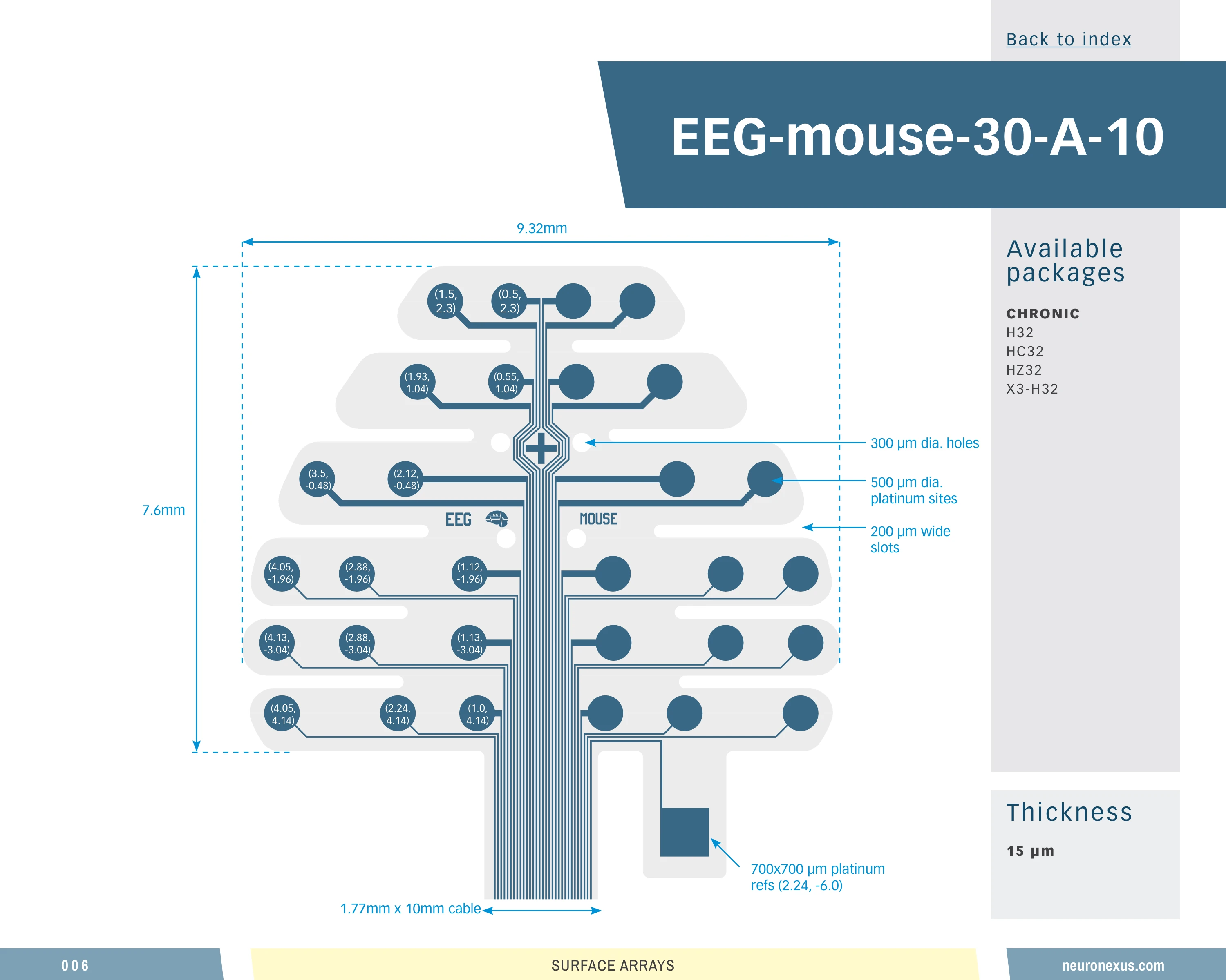
Figure 2. The 30-site mouse EEG layout, drawn to the mouse skull and referenced to bregma, on a 10 mm cable.
The worked example: the 30-site mouse grid on an H32 package#
The 30-site mouse EEG grid, catalog EEG-M-30-A-CL10, on an H32 Hybrid package is the configuration behind the mouse EEG biomarker work of the Binder and Razak laboratories at the University of California, Riverside. Representative examples include the reusable 30-channel technique for awake, freely moving mice of Jonak et al. (2018) ↗, the resting and stimulus-evoked EEG biomarkers in the Fmr1 knockout mouse of Jonak et al. (2020) ↗, and the same biomarkers followed from development into adulthood in Jonak et al. (2024) ↗. The rat layouts of the same series carried the month-long SYNGAP1 study of Katsanevaki et al. (2024) ↗ and Buller-Peralta et al. (2022) ↗, described in Skull-Surface EEG in Rats with a Thin-Film Grid. Further examples are in the NeuroNexus citation database.
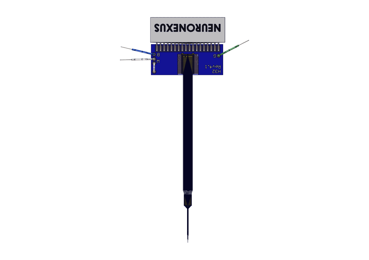
Figure 3. The H32 Hybrid package the grid terminates in: a flexible polyimide cable to an Omnetics 32-pin connector.
How is it implanted and referenced?#
The implant is the one in the EEG surgical guide: screws first, clear of the grid outline, for anchoring and for ground and reference; the bone dried before placement, because the array couples through the skull and a film of fluid under it costs signal; the crosshair on bregma; cement in thin layers over the edges and the cable exit. Impedance that rises over weeks is cement lifting at an edge. The reference decides what a montage means, and the choices, a screw away from the recording area, a grid site, or an external reference, are worked through in Thin Film Grids: EEG Referencing Techniques. Handling and cleaning are in Thin-Film Grid Electrodes: Setup, Handling, Cleaning and Care.
What to order#
The mouse layouts of the EEG Series are the 16-site grid (EEG-M-16-CL10, on an H16 package), the 30-site grid on a 7, 8 or 10 mm cable (EEG-M-30-A-CL7, -CL8, -CL10) and the 32-site grid (EEG-M-32-B-CL10-RET), the 30- and 32-site grids on an H32 Hybrid package that mates a 32-channel headstage; the same preparation runs on XDAQ recording systems with Radiens for acquisition and analysis. The mouse EEG electrode entry has the facts in one place. Talk to an Application Scientist about matching the package to an existing rig, or request a quote with the grid and package names.
Related documents#
- Skull-Surface EEG in Rats with a Thin-Film Grid
- Surgical Guide: Thin Film Grids - EEG Implantation
- Thin Film Grids: EEG Referencing Techniques
- Thin-Film Grid Electrodes: Setup, Handling, Cleaning and Care
- Choosing a Probe Package
Citations#
- Jonak, C. R., Lovelace, J. W., Ethell, I. M., Razak, K. A., and Binder, D. K. (2018). Reusable multielectrode array technique for electroencephalography in awake freely moving mice. Frontiers in Integrative Neuroscience 12, 53. 10.3389/fnint.2018.00053 ↗
- Jonak, C. R., Lovelace, J. W., Ethell, I. M., Razak, K. A., and Binder, D. K. (2020). Multielectrode array analysis of EEG biomarkers in a mouse model of Fragile X Syndrome. Neurobiology of Disease 138, 104794. 10.1016/j.nbd.2020.104794 ↗
- Jonak, C. R., Assad, S. A., Garcia, T. A., Sandhu, M. S., Rumschlag, J. A., Razak, K. A., and Binder, D. K. (2024). Phenotypic analysis of multielectrode array EEG biomarkers in developing and adult male Fmr1 KO mice. Neurobiology of Disease 195, 106496. 10.1016/j.nbd.2024.106496 ↗
- Katsanevaki, D., et al. (2024). Key roles of C2/GAP domains in SYNGAP1-related pathophysiology. Cell Reports 43, 114733. 10.1016/j.celrep.2024.114733 ↗
- Buller-Peralta, I., et al. (2022). Abnormal brain state distribution and network connectivity in a SYNGAP1 rat model. Brain Communications 4(6), fcac263. 10.1093/braincomms/fcac263 ↗