Skull-Surface EEG in Rats with a Thin-Film Grid
A 32-channel polyimide grid on the rat skull records high-density EEG for a month with no craniotomy, resolving state-dependent biomarkers in a SYNGAP1 model.
4 min read
Updated September 23, 2026
A 32-channel polyimide thin-film grid, aligned to bregma on the intact rat skull, recorded high-density EEG for up to one month with no craniotomy, in six-hour sessions at 1 kHz with EMG and accelerometry from the same implant. It resolved altered sleep-wake distributions and reduced inter-regional coherence in SYNGAP1 heterozygous rats — state-dependent network differences that map onto the constructs used in human high-density EEG.
Research use only. NeuroNexus products and the procedures described here are for non-clinical research use, and must not be used in human or veterinary medical procedures.
Can rodent EEG be recorded densely enough, and for long enough, to serve as a translational biomarker?#
That was the question. Human studies of neurodevelopmental disorders use high-density scalp EEG — power by band and by region, connectivity between regions, the distribution of brain states across the day — and a rodent model is only useful as a bridge if the same constructs can be measured in it, over the weeks a phenotype takes to express, without the surgery that changes the animal. Single screw electrodes give too few channels for a connectivity claim; a cortical-surface array gives more channels at the cost of a craniotomy and a preparation that scars. The requirement was dense coverage on the skull surface, held stable for a month, with placement comparable between animals.
How the recording was made#
The array is a 12 µm polyimide thin-film grid ↗ from the EEG series ↗, the rat functional layout EEG-R-32-A-CL10-func, with an integrated polyimide ribbon cable and 500 µm platinum sites. Fourteen sites recorded EEG; two further channels connected to bilateral EMG leads — 6.5 cm stainless steel, Teflon-coated — so that sleep and wake staging came from the same implant as the EEG. The grid terminated in an H32 Hybrid package ↗ on an Omnetics connector.
Placement is the experiment on the skull. A crosshair fiducial on the grid was aligned to bregma ↗ and the array anchored with no bone removed at any point, which is what makes site positions comparable between subjects; the procedure is the one in the EEG surgical guide. Sessions ran six hours at 1 kHz inside a 1 × 1 m Faraday cage on an Open Ephys system with a 32-channel headstage and an integrated accelerometer. Adult rats were followed for up to one month.

Figure 1. The rat functional thin-film EEG grid: 12 µm polyimide, 500 µm platinum sites laid out to the rat skull, with the bregma crosshair.
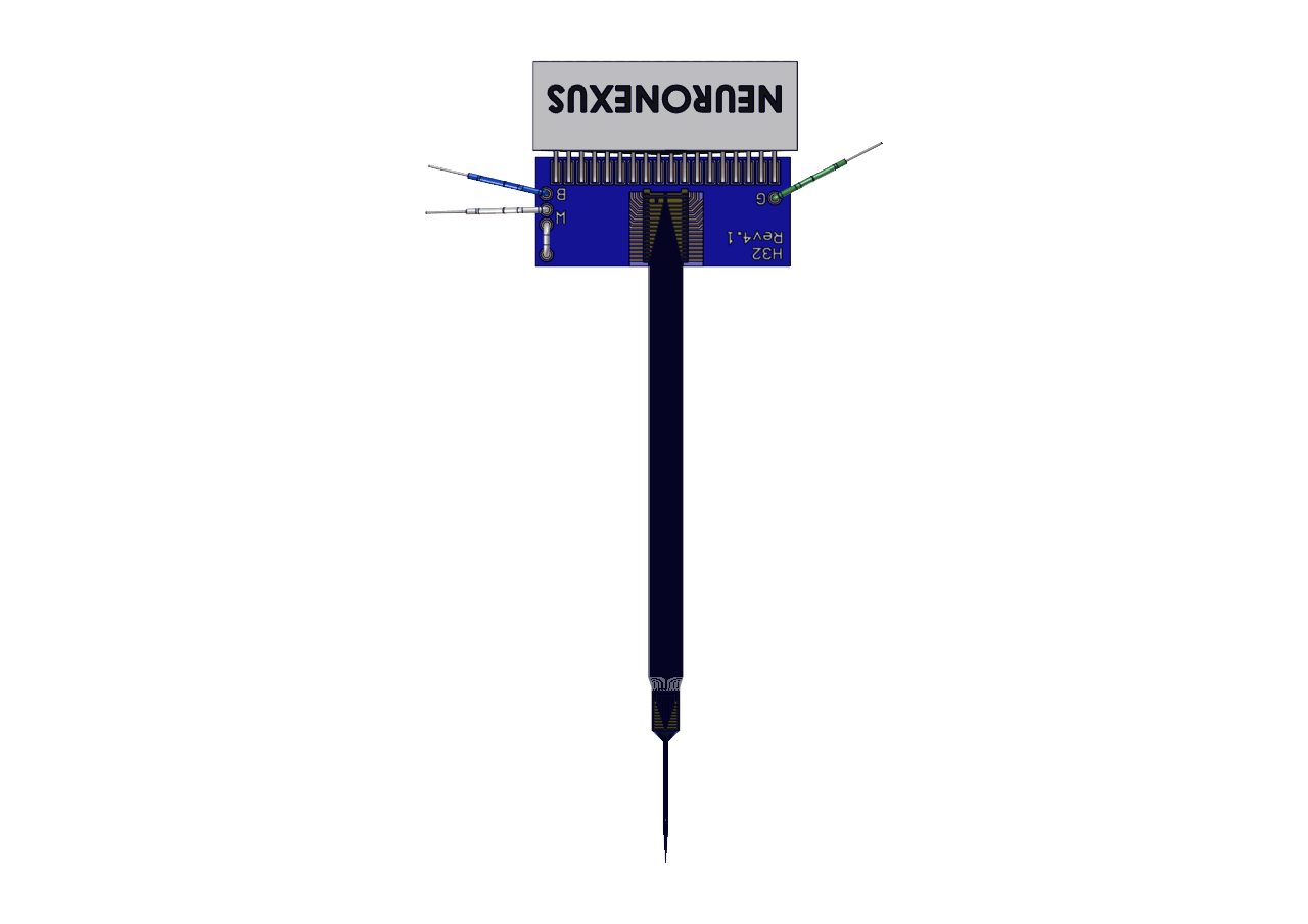
Figure 2. The H32 Hybrid package the grid terminates in: a flexible polyimide cable to an Omnetics 32-pin connector.
What the grid resolved#
State-specific spectral and connectivity differences, in vivo, across sessions. Sleep-wake distributions shifted in SYNGAP1 heterozygotes relative to wild-type littermates. Band-limited power differences localized over frontal and parietal channels and depended on state. Magnitude-squared coherence between regions fell in defined frequency bands, and the effect persisted across sessions over the month. Staging came from the EMG and the accelerometer rather than from the EEG being analyzed, so state and spectrum were measured on the same clock without circularity. The full analyses are in the two papers cited below.
What it means for the method#
Three things held the effects stable across subjects, and they transfer to any power or coherence workflow: bregma alignment, so that a channel means the same region in every animal; a fixed six-hour session structure at fixed acquisition parameters; and staging from an independent signal on the same implant. The measurements map onto constructs used in human HD-EEG analysis, and skull-surface placement preserves the volume-conduction path that makes the comparison meaningful — a cortical-surface array would record more, but not the same thing. What the skull costs is spatial resolution: a result that depends on a fine spatial claim needs an ECoG grid instead, and the reference decision, which determines what a montage means, is worked through in Thin Film Grids: EEG Referencing.
What to order#
The array is the catalog EEG-R-32-A-CL10-func grid on an H32 Hybrid package; the mouse layouts and the other rat layouts in the EEG series ↗ follow the same procedure. The same preparation runs on XDAQ recording systems with Radiens for acquisition and analysis, and the placement procedure is described in the investigators' papers, which makes cross-site replication practical. Talk to an application scientist about matching the package to an existing rig, or request a quote ↗ with the grid and package names.
Publications. Katsanevaki et al., 2024, Cell Reports 43, 114733 ↗ · Buller-Peralta et al., 2022, Brain Communications 4(6), fcac263 ↗.