NeuroNexus Probe Technology: Questions and Answers
Questions on NeuroNexus silicon probes and thin-film grids, answered from the published record: lineage, thickness, dielectrics, SIROF, Z-Coat, chronic use.
11 min read
Updated October 27, 2026

NeuroNexus · neuronexus.com · Resources
NeuroNexus combines the heritage of the original Michigan Probe (Wise et al. (1970) ↗; Drake et al. (1988) ↗) with advanced materials, proprietary microfabrication processes, and continuous innovation to produce robust, reproducible silicon neural probes for electrophysiology research. NeuroNexus probe designs support neural recording, stimulation, and multi-modal optogenetics across experimental models ranging from organoids and fruit flies to non-human primates, and applications ranging from intra-operative, acute studies to implanted, chronic studies.
Research use only. NeuroNexus products are for non-clinical research use and must not be used in human or veterinary medical procedures.
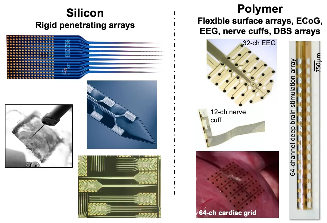
Figure 1. The two NeuroNexus thin-film electrode platforms: silicon probes for penetrating targets (left) and polymer grids for surface targets (right).
How has NeuroNexus advanced the original Michigan probe?#
NeuroNexus spun out of the University of Michigan in 2004, and its current leaders and founders (Hetke, Vetter, and Kipke) were members of the Michigan probe development team. Over the past twenty years, NeuroNexus has continually improved probe components and materials as the field has advanced, including the silicon substrate, dielectric (insulating) layers, metal interconnects, electrode sites, and packages. It has also developed hundreds of purpose-driven probe designs that today provide the most extensive catalog of probes and packages, targeting almost any site in the brain, spinal cord, periphery, and heart. What began as an academic development program at Michigan, funded by the NIH, is now a mature commercial product platform used by researchers worldwide. Additional history of silicon probe development is in A Brief History of Electrode Technology.
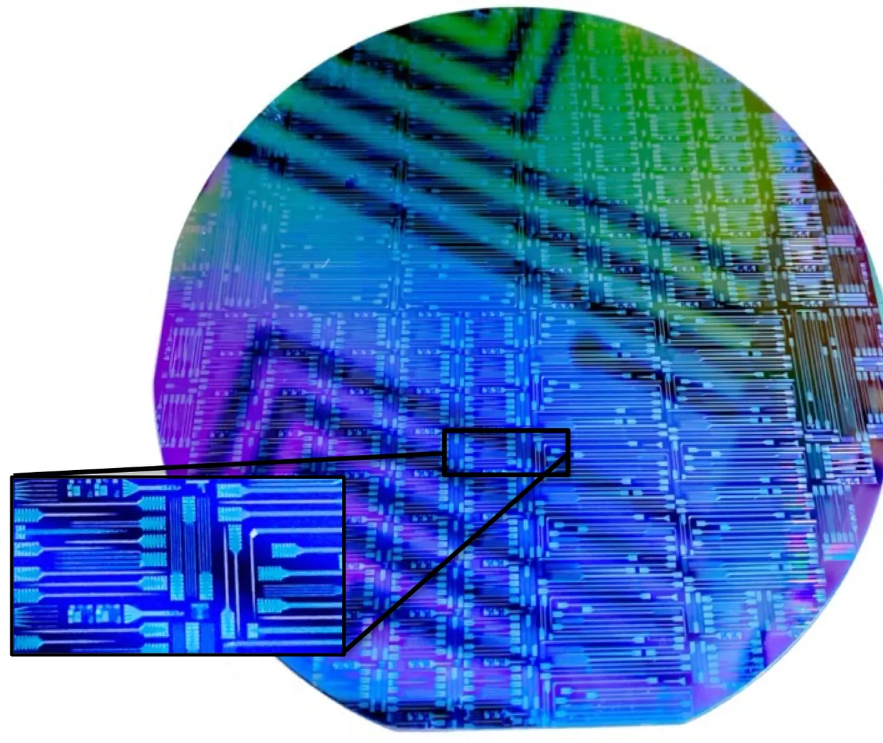
Figure 2. A silicon wafer of microfabricated NeuroNexus probes before release, with an inset of individual shanks and bond pads.
Why does silicon probe thickness matter?#
Substrate thickness influences bending stiffness, tissue displacement, handling, and suitability for accessing different recording depths. NeuroNexus offers silicon probes in 5, 15, and 50 µm thicknesses to match specific experimental requirements. Ultra-thin designs (5 µm) offer greater flexibility and can reduce tissue displacement (Seymour and Kipke (2007) ↗), while 15 and 50 µm substrates offer more stiffness. For larger animal models and deeper insertions into deep brain regions, additional thicker or mechanically reinforced designs are available, including 110mm-long Vector probes. In general, probe selection for a particular target and application involves considering target anatomy, probe geometry, surgical techniques, and experimental functional requirements.
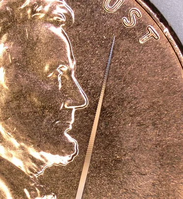
Figure 3. A Vector probe tip beside a US penny: a short silicon array on a rigid support body for deep targets in large animals.
Why does NeuroNexus use amorphous silicon carbide dielectrics?#
NeuroNexus uses a proprietary low-stress thin-film amorphous silicon carbide (a-SiC) as the insulating (dielectric) layer, for electrical isolation and chemical stability in tissue, including for long-term chronic use. The performance of a-SiC is well documented. Cogan et al. (2003) evaluated plasma-deposited silicon carbide as an implantable insulating coating and reported excellent insulation and stability under physiological conditions, and Deku et al. (2018) built a-SiC ultramicroelectrode arrays for recording and stimulation.
How are conductive traces engineered for reliable performance?#
NeuroNexus uses a proprietary multilayer metal stack that connects electrode sites to bond pads, optimized for feature size, adhesion, electrical resistance, and manufacturability. This metallization is part of a tightly controlled microfabrication process engineered to deliver consistent device characteristics from design through production.
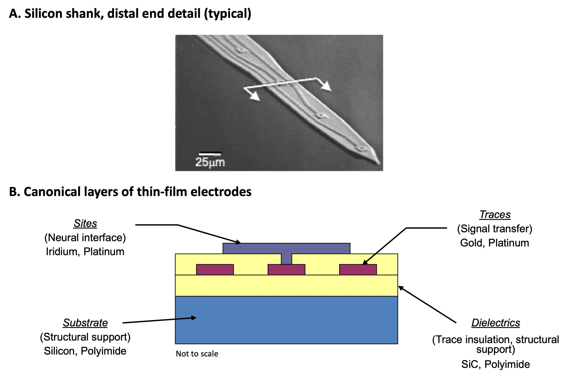
Figure 4. Thin-film electrode construction. (A) Electron micrograph of a silicon shank tip with its electrode sites. (B) The layer stack: substrate, dielectric (insulating) layers, metal traces, and electrode sites.
Why are sputtered iridium oxide film (SiROF) electrode sites useful for recording and stimulation?#
Sputtered iridium oxide film (SIROF) electrode sites provide low impedance, typically below 100 kOhm for representative NeuroNexus configurations, and high charge-injection capability for electrical stimulation. Actual impedance depends on site geometry and measurement conditions Cogan (2008) ↗, Cogan et al. (2009) ↗.
How does Z-Coat (PEDOT) further reduce electrode impedance?#
Z-Coat is NeuroNexus's proprietary PEDOT site coating that lowers site impedance by roughly an order of magnitude compared with an uncoated site of the same area and increases charge injection capacity, without changing the site footprint. More information on the Z-coat coating, its stability, and its use for stimulation is in Z-Coat: Optimized Site Coating for Stimulation and Noise Reduction.
Can NeuroNexus probes be used in acute and chronic studies?#
Yes. NeuroNexus silicon probes are used in both acute (intraoperative) and chronic studies. The same silicon probes are used for both acute and chronic studies. Researchers typically select the probe package for specific acute or chronic applications. Select examples include: Okun et al. (2016) reported long-term recordings with immobile silicon probes in mouse cortex; Haghighi et al. (2025) implanted multishank silicon probes in rat somatosensory cortex for 16 weeks; Mueller et al. (2025) used a 16-site NeuroNexus probe as the silicon comparator in a 12-week chronic recording study in rat.
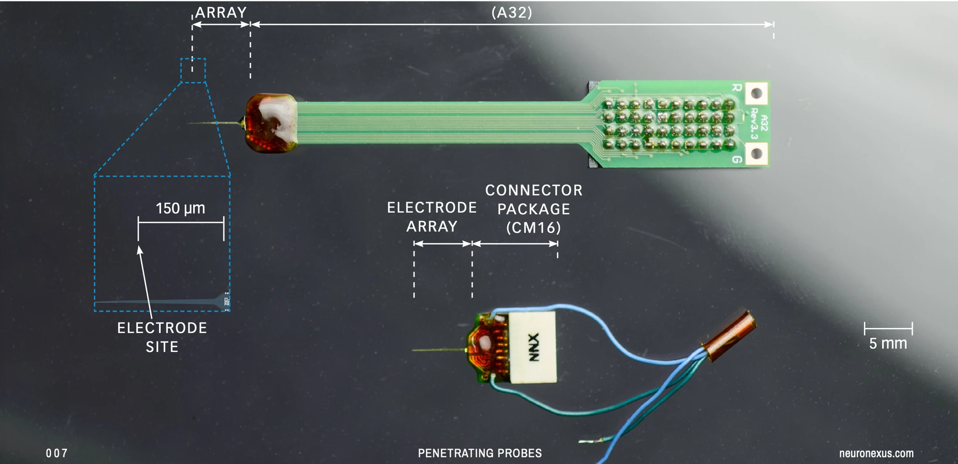
Figure 5. A probe assembly: the electrode array joined to its connector package, on an A32 package (top) and a CM16 package (bottom). The package is selected for the acute or chronic application.
Can NeuroNexus probes be used in diverse experimental preparations, from organoids to non-human primates?#
Yes. NeuroNexus offers a broad selection of silicon neural probes with electrode geometries, channel counts, shank lengths, and substrate thicknesses designed to accommodate diverse animal models, from fruit flies to non-human primates.
NeuroNexus technology's versatility is reflected in more than 5,100 citations in scientific publications across species, brain regions, and experimental approaches. Representative examples include Van De Poll and van Swinderen (2025) ↗, who used NeuroNexus probes to investigate sleep and wakefulness in fruit flies, and Thirunavukkarasu et al. (2026) ↗, who used NeuroNexus laminar probes to examine sensory responses in macaques. In another recent non-human primate study, Perich et al. (2026) ↗ used NeuroNexus Vector arrays to record neural activity from deep brain structures. Vetter et al. (2004) ↗ first reported chronic performance using silicon probes for intracortical recording of spikes and fields.
These papers illustrate just a small fraction of the applications enabled by NeuroNexus probes. Explore many more examples in the NeuroNexus scientific publication database.
How broad is the silicon probe catalog and custom offerings?#
NeuroNexus offers a silicon probe catalog with over 210 shank-geometry varieties, electrode-site arrangements, channel counts, lengths, and thicknesses. Researchers can select from established catalog configurations or work with NeuroNexus on custom probe design and fabrication, including specialized geometry and packaging requirements.
How do NeuroNexus thin-film polymer grid electrode arrays relate to silicon probes?#
NeuroNexus thin-film polymer grids are the surface counterpart of its silicon probes, made by the same proprietary thin-film microfabrication process on a flexible 12 µm polyimide substrate rather than rigid silicon. A grid rests on the surface of the cortex, skull, heart, or a peripheral nerve and records the field potential beneath each site, where a silicon probe penetrates the tissue and records at depth. The catalog includes 53 grid designs in four families, ECoG, EEG, cardiac surface, and peripheral nerve cuff, with platinum sites from 25 µm to 1 mm in diameter and 3 to 256 sites per array, and NeuroNexus fabricates custom footprints, site layouts, and cable lengths on the same process. Grids connect to the same recording chain of headstages and acquisition systems as silicon probes, and researchers routinely combine the two in one preparation to record surface and depth together. The grid families, their placement, and site-size selection are described in Thin-Film Grids for Surface Recording.
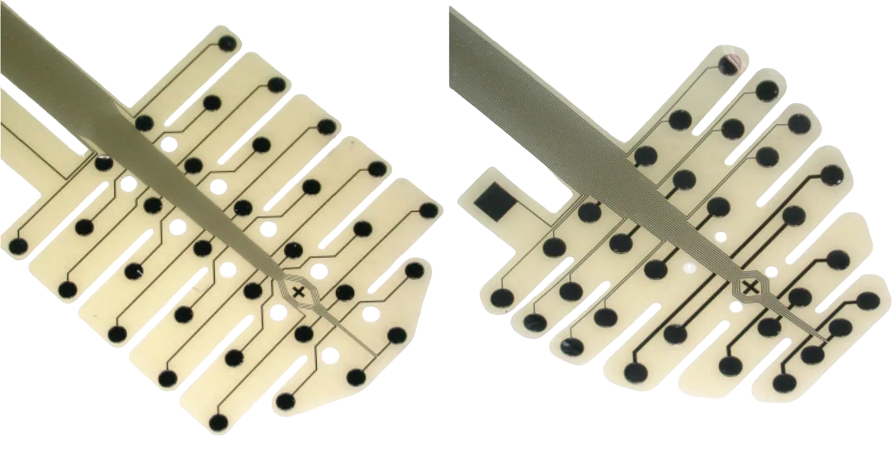
Figure 6. Thin-film polymer EEG grids for the mouse skull, in two layouts, on the 12 µm polyimide substrate shared by every NeuroNexus grid.
How widely has NeuroNexus technology been used in published research?#
NeuroNexus technology has appeared in more than 5,000 peer-reviewed publications across neuroscience and bioengineering research. See the NeuroNexus citations resource ↗ for publications and product-family citation information. Citation counts reflect the company’s broader product portfolio, not just current-generation silicon probes.
Related documents#
- Read next, in order: Silicon Probes and Thin-Film Grids: An Introduction · How an Electrode Records Neural Activity · What Makes a Thin-Film Electrode Good · Choosing an Electrode for Your Experiment · Choosing a Probe Package · Thin-Film Grids for Surface Recording · The Electrode Design Space
- Materials and care: Z-Coat: Optimized Site Coating for Stimulation and Noise Reduction · Silicon Probes: Setup, Handling, Cleaning and Care · A Brief History of Electrode Technology
- Glossary: Silicon probe · Thin-film grid · Substrate thickness · Site area · Impedance · Probe package · Thin-film microfabrication
Citations#
- Vetter, R. J., et al. (2004). Chronic neural recording using silicon-substrate microelectrode arrays implanted in cerebral cortex. IEEE Transactions on Biomedical Engineering, 51(6), 896–904. DOI: 10.1109/TBME.2004.826680 ↗
- Cogan, S. F., et al. (2003). Plasma-enhanced chemical vapor deposited silicon carbide as an implantable dielectric coating. Journal of Biomedical Materials Research Part A, 67(3), 856–867. DOI: 10.1002/jbm.a.10152 ↗
- Deku, F., et al. (2018). Amorphous silicon carbide ultramicroelectrode arrays for neural stimulation and recording. Journal of Neural Engineering, 15(1), 016007. DOI: 10.1088/1741-2552/aa8f8b ↗
- Cogan, S. F. (2008). Neural stimulation and recording electrodes. Annual Review of Biomedical Engineering, 10, 275–309. DOI: 10.1146/annurev.bioeng.10.061807.160518 ↗
- Cogan, S. F., et al. (2009). Sputtered iridium oxide films for neural stimulation electrodes. Journal of Biomedical Materials Research Part B, 89(2), 353–361. DOI: 10.1002/jbm.b.31223 ↗
- Okun, M., et al. (2016). Long term recordings with immobile silicon probes in the mouse cortex. PLOS ONE, 11(3), e0151180. DOI: 10.1371/journal.pone.0151180 ↗
- Mendoza, G., et al. (2016). Recording extracellular neural activity in the behaving monkey using a semichronic and high-density electrode system. Journal of Neurophysiology, 116(2), 563–574. DOI: 10.1152/jn.00116.2016 ↗
- Paulk, A. C., et al. (2013). Multichannel brain recordings in behaving Drosophila reveal oscillatory activity and local coherence in response to sensory stimulation and circuit activation. Journal of Neurophysiology, 110(7), 1703–1721. DOI: 10.1152/jn.00414.2013 ↗
- Van De Poll, M. N., & van Swinderen, B. (2025). Long-term multichannel recordings in Drosophila flies reveal altered predictive processing during sleep compared with wake. Journal of Experimental Biology, 228(11), jeb250165. DOI: 10.1242/jeb.250165 ↗
- Perich, M. G., et al. (2026). Inferring brain-wide interactions using data-constrained recurrent neural network models. Neuron. DOI: 10.1016/j.neuron.2026.07.016 ↗
- Thirunavukkarasu, P., et al. (2026). Laminar architecture of visual and auditory responses in the supplementary eye field of macaques. Cerebral Cortex, 36(6), bhag064. DOI: 10.1093/cercor/bhag064 ↗
- Haghighi, et al. (2025). Piezo1 and Piezo2 Ion Channels in Neuronal and Astrocytic Responses to MEA Implants in the Rat Somatosensory Cortex. International Journal of Molecular Sciences. DOI: 10.3390/ijms26189001 ↗
- Mueller, et al. (2025). Mechanically-adaptive, resveratrol-eluting neural probes for improved intracortical recording performance and stability. npj Flexible Electronics. DOI: 10.1038/s41528-025-00440-5 ↗
- Wise, K. D., Angell, J. B., & Starr, A. (1970). An integrated-circuit approach to extracellular microelectrodes. IEEE Transactions on Biomedical Engineering, 17(3), 238–247. DOI: 10.1109/TBME.1970.4502738 ↗
- Drake, K. L., Wise, K. D., Farraye, J., Anderson, D. J., & BeMent, S. L. (1988). Performance of planar multisite microprobes in recording extracellular single-unit intracortical activity. IEEE Transactions on Biomedical Engineering, 35(9), 719–732. DOI: 10.1109/10.7273 ↗
- Seymour, J. P., & Kipke, D. R. (2007). Neural probe design for reduced tissue encapsulation in CNS. Biomaterials, 28(25), 3594–3607. DOI: 10.1016/j.biomaterials.2007.03.024 ↗